
Contact Admission
International Collaboration


Identifying Collagen Vascular Disease Through Persistent Skin Lesions
A case report published in JAMA Dermatology highlights a rare condition known as Cutaneous Collagenous Vasculopathy (CCV), which was diagnosed after the patient had lived with unexplained purpuric lesions for more than 20 years.
Symptoms of Cutaneous Collagenous Vasculopathy: Clues From Persistent Purpuric Macules
The clinical case involves a 60-year-old male patient with a history of hypertension, hyperlipidemia, and coronary artery disease, who presented with multiple asymptomatic purpuric patches that had persisted for over two decades. The lesions initially appeared on both lower legs and gradually spread to the inner thighs and then the forearms.
Symptoms worsened with prolonged standing and were occasionally accompanied by mild pruritus. On examination, the patient exhibited diffuse, reticular telangiectatic patches that were blanchable and symmetrically distributed over the legs and forearms. Dermoscopic examination revealed fine, blanchable telangiectasias. Notably, there were no mucosal or nail abnormalities, no family history of similar conditions, and no signs of systemic disease or coagulopathy.
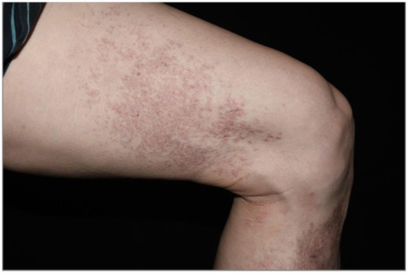
Diffuse, reticular, blanchable telangiectatic patches symmetrically covering the legs. Image: JAMA Dermatology
To determine the underlying cause, a skin biopsy was performed on the patient’s left lower leg. Histopathologic examination revealed dilation of small blood vessels in the upper dermis, with concentric deposition of amorphous, pink, hyaline-like material within the vessel walls.
This material tested positive with periodic acid–Schiff (PAS) staining but negative with Congo red, ruling out amyloidosis. Based on these findings, the patient was diagnosed with Cutaneous Collagenous Vasculopathy (CCV). As there were no systemic symptoms or involvement of internal organs, he was advised to undergo regular monitoring. The lesions remained stable after 3 months of follow-up.
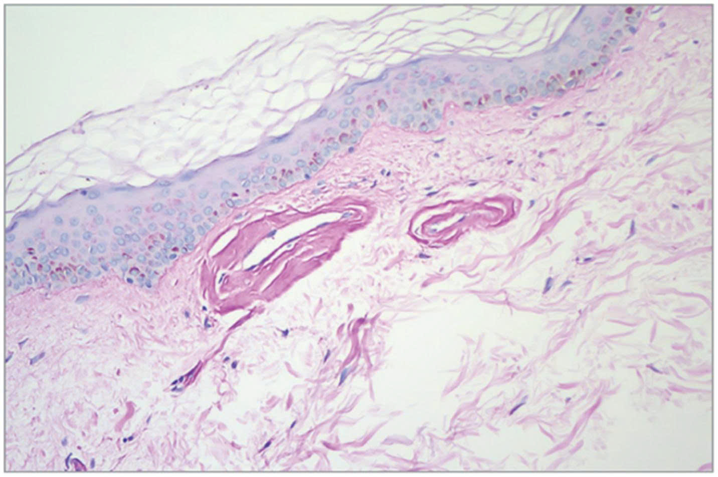
Histologic examination shows dilated small vessels in the upper dermis with concentric deposition of amorphous pink hyaline-like material within the vessel walls. Image: JAMA Dermatology.
What Is CCV and How Is It Treated?
Cutaneous Collagenous Vasculopathy (CCV) is a rare microvascular disorder that affects superficial blood vessels just beneath the skin. Clinically, it presents as painless telangiectatic patches or tiny purpuric macules, most commonly on the lower extremities, with a tendency to progress upward. CCV typically affects middle-aged and older adults, with no significant difference in prevalence between men and women.
Pathogenesis and Mechanism The exact cause of CCV remains unclear, but researchers suggest that mild, chronic endothelial injury may lead to fibrin-like hyaline material deposition in vessel walls. Over time, this accumulation results in fibrosis surrounding the blood vessels, contributing to the characteristic histological features of the disease.
Management and Treatment Options As CCV rarely involves internal organs, management is generally conservative and focused on cosmetic improvement if requested by the patient. The most effective intervention currently reported is pulsed dye laser therapy, which can reduce the appearance of telangiectatic lesions.
Clinical Significance and Diagnostic Considerations Though rare, CCV presents with distinct clinical and histopathologic features. Recognizing its characteristic signs and confirming the diagnosis via skin biopsy is key to distinguishing it from other vascular dermatoses and ensuring accurate management.
Differential Diagnosis: How to Distinguish CCV From Similar Conditions
Diagnosing CCV relies heavily on histopathologic examination, as it may clinically resemble several other telangiectatic or purpuric skin disorders:
Generalized Essential Telangiectasia: Also features symmetric telangiectatic macules on the lower limbs and body, but biopsy typically shows only mild capillary dilation without the hyaline deposits characteristic of CCV.
Schamberg’s Disease: Presents as asymptomatic brownish macules or petechiae on the lower extremities with a classic “cayenne pepper” appearance. Histology reveals perivascular inflammation, erythrocyte extravasation, and hemosiderin-laden macrophages.
Hereditary Hemorrhagic Telangiectasia (HHT): A genetic disorder featuring multiple arteriovenous malformations (AVMs) typically on the hands, face, lips, tongue, and oral mucosa. Unlike CCV, HHT often involves a positive family history of abnormal bleeding (e.g., recurrent nosebleeds) and may affect visceral organs such as the GI tract or lungs.
Conclusion CCV is a rare but distinct vascular disorder with unique clinical and histopathologic features. Proper identification of its hallmark signs and accurate biopsy techniques are essential to differentiate it from similar dermatologic conditions, enabling appropriate diagnosis and patient care.
If you or someone you know develops persistent, asymptomatic red or telangiectatic patches, particularly on the legs, it's advisable to consult a dermatologist for proper evaluation and timely diagnosis.
Read the full article as published in JAMA Dermatology here
Other Case Report
- Viêm da tiết bã, dấu hiệu rối loạn hàng rào biểu mô hệ thống ( 07:44 - 08/11/2025 )
- Artificial Intelligence in Surgery ( 14:48 - 06/09/2025 )
- Abrocitinib: A New Direction in the Treatment of SAPHO Syndrome ( 14:49 - 20/06/2025 )
- MỘT TRƯỜNG HỢP ABSCESS VÙNG HANG VỊ- CĂN BỆNH HIẾM GẶP- ĐƯỢC ĐIỀU TRỊ THÀNH CÔNG TẠI BỆNH VIỆN TÂM TRÍ CAO LÃNH ( 09:34 - 09/05/2025 )
- Phẫu thuật Mitrofanoff: 40 năm nhìn lại. ( 08:35 - 11/04/2025 )
- SỬ DỤNG MẬT ONG HOA TRÀM CHIẾU XẠ TRONG ĐIỀU TRỊ LOÉT TÌ ĐÈ ( 08:21 - 28/03/2025 )
- Chronic Progressive Pink-Yellow Papules and Nodules in a Middle-Aged Man ( 09:02 - 24/09/2024 )
PHAN CHAU TRINHUNIVERSITY
- 09 Nguyen Gia Thieu street,
Dien Ban Dong ward,
Da Nang city
Phan Chau TrinhUniversity’s Hospital
- 09 Nguyen Gia Thieu street,
Dien Ban Dong ward,
Da Nang city