
Contact Admission
International Collaboration


To be daring
More than 350 years ago, an English natural philosopher - Robert Hooke looked through a microscope at a small node and discovered that "it" was made from small box-like compartments, which he named "the sacrifice planing ”. From that moment, Hooke and countless curious minds after he tried to have a better view of the basic building blocks of this life.
Thiết bị kính hiển vi mới chụp các bộ phim 3D những tế bào bên trong các sinh vật sống một cách chi tiết chưa từng có.
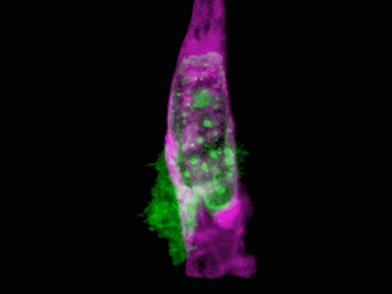
In a new study in the April 20 issue of Science, researchers from Howard Hughes Medical Institute's Janelia Institute of Medicine (HHMI), Harvard Medical School and collaborative organizations report The development of a microscope capable of 3D imaging and the cells inside an organism's life in unprecedented detail.
In accordance with the technique used by astronomers to study distant stars, the team, led by Nobel Prize winner and team leader Janelia Eric Betzig, introduced this new technology by creating A series of great films like: Cancer cells crawling through the blood vessels, Spinal nerve cells inserted into the vessels, Immune cells that fly into the ears of zebrafish and much more. The microscope's resolution is strong enough to capture details in a cellular form, such as the dynamics of tiny bubbles called blisters, which transport molecular goods across cells. Study co-author Tomas Kirchhausen - Harvard Medical School cell biology professor - Springer Family President of Pediatrics and a senior investigator at Children's Hospital Boston said: “It's a miracle to be able to see See what we've never seen before. It was amazing ”. “Every time we do an experiment with this microscope, we see something new - and create new ideas and hypotheses to test,” says Kirchhausen. "The microscope can be used to study any problem in a biological system or organism that I think of."
While scientists have used the microscope to observe cells for centuries, the most obvious views ever come from cells isolated on glass slides. Visualizing real-time living cells inside living organisms is still much more difficult. Cells surrounded by tissues and other biological structures scramble for light coming from and back to a microscope target, blurring and obscuring important details. Light is strong enough to penetrate biological structure and give a sharper view of cells, on the other hand, can damage tissues.
"This raises suspicions that we don't see peaceful natural cells in the organism in which they evolved," said Betzig, co-author of the study. It is often said that seeing is believing, but when it comes to cell biology, I think the more appropriate question is: "When can we believe what we see?" To address these challenges, Betzig et al. Combined two technologies: the optical microscope, Betzig developed in the early 2010s, and adaptive optics, a technique borrowed from astronomy. . The optical microscope scans quickly and repeatedly of an ultra-thin sheet of light, helping to avoid bleaching or damage associated with the traditional focused light beam. It can be used to produce high-resolution 2D images of living cells as they perform functions, and by combining these series of images over time, scientists can create 3D movies. To rearrange the plate of light as it passes through other tissues and structures, the team turned to the stars. To view distant objects through the Earth's atmosphere, astronomers rely on adaptive optics - strain mirrors and light modulators. This process uses a powerful laser, aimed at the small sky space they want to image, serving as a "guide star". As the laser passes through the atmosphere, the optical aberrations that distort its path are exposed and corrected by adaptive optics. Betzig and his colleagues applied this principle to the microscopic world, using a two-photon laser to create an adaptive optical system that maintains the thin illumination of the slab of light as it passes through an organism to creates undistorted images.
The team confirmed the new optical microscope on a series of biological samples, mostly done through the laboratories of Kirchhausen and Sean Megason, professor of biology at Harvard Medical School. To understand the data they generated, the team created separate software and computational and visual workflows, led by co-study author Gokul Upadhyayula, a professor at Harvard Medical School in Pediatrics. in Boston and research associates Kirchhausen and Tsung-Li Liu, formerly a research scientist at the Betzig Laboratory at the Howard Hughes Medical Institute. "For the types of data we generate, there is no commercial software we can use to make movies that can interpret and extract biologically meaningful information, because So we built the necessary tools.This allows us to understand what we collect and visualize data in in meaningful ways, including, in the near future, interactive 3D movies. full".
K
The results were remarkable. In one movie, a fiery orange immune cell giggles frantically through a zebrafish's ear while scooping green sugar particles along the way. In another, a metastatic cancer cell attaches to its attached appendages as it rolls through a blood vessel and tries to squeeze the vessel wall. The team collected films about organelles' behavior as they self-repair inside cells during cell division and were even able to visualize in real time and near the genus. protein-mediated intracellular secretion of clathrin, which cells use to capture materials from their external environment. "I work to understand how 'eating' cells use machines based on carriers," says Kirchhausen, and all my life I have dreamed of this in a living organism. We have finally achieved this ”. The complexity of 3D multicellular media can be overwhelming, Betzig said, but the clarity of the image allows them to compute "explosions" in addition to individual cells in the tissue to focus on internal dynamics. any particular one. "We don't even know the questions to ask because we have never seen some of these organisms at this level of detail," Upadhyayula said. without adaptive optics, Betzig said, "it's too faint." In his view, adaptive optics is one of the most important fields in microscope research today, and microscopy. Learning, excels at 3D live visuals, is the perfect platform for displaying its power.
The next step is to make the technology affordable and user-friendly. The current microscope occupies a table 10 feet long. In collaboration with Kirchhausen and Upadhyayula, Betzig's team is working on a next-generation version that should fit a small table at a cost within reach of individual laboratories. Such tool will first go to the Janelia Advanced Imaging Center, where scientists from around the world can apply to use it. A second device built at the same time will be located at the Kirchhausen laboratory at Harvard Medical School in Boston. A tool building plan will also be provided.
Contributing authors include researchers from Stony Brook University, University of California, Berkeley, California Institute of Technology and University of Exeter.
Study supported by Howard Hughes Medical Institute, National Institutes of Health (R01GM075252, R01DC015478, 5R00CA154870-05, 1R01GM121597-01, R01CA196884 and R35GM118149), National Science Foundation (IOS1452928), Breast Cancer Research Foundation Carol M. Baldwin, Damon Runyon Cancer Research Foundation, Pew Charitable Foundation, Biogen, Ionis Pharmaceuticals and Frontier Human Science Program.
Adapted from a Howard Hughes Medical Institute newsletter.
Source:https://hms.harvard.edu/news/boldly-go?utm_source=Silverpop&utm_medium=email&utm_term=s1&utm_content=4.23.18.HMS
Translation summary: Dr. Nguyen Huu Tung et al
Other news
- How Dangerous Is Nipah Virus? Medical Alert and Urgent Health Recommendations ( 14:13 - 27/01/2026 )
- Predicting Disease from Sleep – A New Breakthrough Study ( 14:01 - 13/01/2026 )
- Medical advances predicted to break through in 2026 ( 13:54 - 12/01/2026 )
- Vietnamese medical miracles in 2025 – inspiration for medical students ( 07:54 - 07/01/2026 )
- Updating the SOFA-2 Score: A New Standard in the Assessment of Multiple Organ Failure After Three Decades ( 10:40 - 31/12/2025 )
- Home AEDs: High Life-Saving Effectiveness, but Not Cost-Effective at Current Prices ( 14:12 - 18/12/2025 )
- Artificial Intelligence and Pediatric Care ( 08:27 - 16/12/2025 )
- Applying Clinical Licensing Principles to Artificial Intelligence ( 09:36 - 08/12/2025 )
- U.S. Approves Targeted Lung Cancer Therapy Datroway ( 08:43 - 25/06/2025 )
- Therapeutic potential and mechanisms of mesenchymal stem cell-derived exosomes as bioactive materials in tendon–bone healing ( 08:38 - 23/11/2023 )
PHAN CHAU TRINHUNIVERSITY
- 09 Nguyen Gia Thieu street,
Dien Ban Dong ward,
Da Nang city
Phan Chau TrinhUniversity’s Hospital
- 09 Nguyen Gia Thieu street,
Dien Ban Dong ward,
Da Nang city